Complications of Blepharoplasty: Prevention and Management
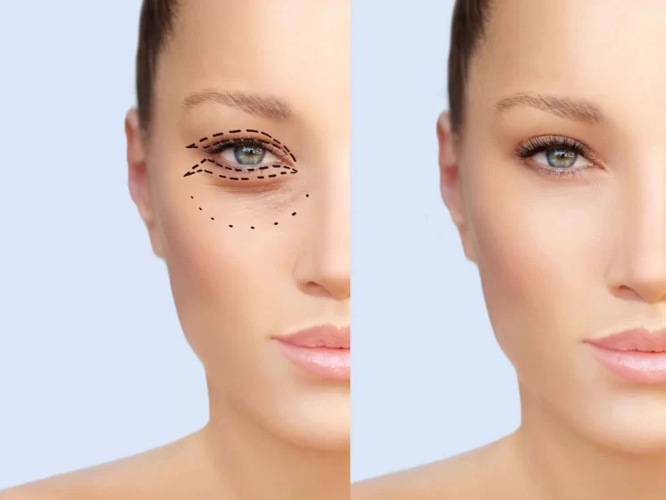
Blepharoplasty commonly called eyelid surgery is an effective procedure in order to remove the droopy skin and puffy bags from upper and lower eyelid. It can also improve the side (peripheral) vision which is hampered through extra skin fat and skin pockets around the eyes. This surgery gives a younger look to your face as eyes are the central feature of the face. It does not serve only aesthetic reasons but medical as well. An expert surgeon will make you aware about all the potential complications which can occur after the surgery. He will certainly try to minimize the same by giving you accurate guidance and preoperative tips. As a patient you must inform about your physical health, medication you are taking, the type of exercise you do and the diet you take. A detailed discussion about your expectations and the possibilities that surgery can offer should be done with the surgeon as you must have real expectation. Generally, almost every surgery has some or little side-effects that get away with time and sometimes there are permanent issues. Eyelid surgery is also the same and carries the risk of bleeding, bruising and infection which are easily manageable if treated by a skilled and expert surgeon. As a patient you must be aware about many other complications which are as follows:-
Some easily manageable risks and complications include:
- blurry vision
- damage from excessive sun exposure
- dry eyes
- itchiness around the eye area
- inability to close your eyes
- muscle damage
- scars
Seek medical attention immediately if you experience any of the following:
- Shortness of breath
- Chest pain
- An unusual heart rate
- Severe new eye pain
- Continuous Bleeding
- Vision problems
There are some complications which can bother you for a lifetime or these will be treated only with revision surgeries, therefore the key to eliminate such issues of blepharoplasty is to avoid them at all costs. This means from the beginning when you take this decision and consult a surgeon: – Do the discussions thoroughly as many times as possible to set realistic goals and have extensive knowledge to overcome the fear of surgery and its complications.
Some of the most unpleasant complications and the ways to avoid them are as follows:-
Infection:
Infection after a blepharoplasty is rare. Usually the patients are put on an antibiotic ointment for the first postoperative week. Late and chronic infection with atypical mycobacteria is a possibility and will present as red, raised nodules along the incisions. Treatment is the appropriate antibiotic. In the case of atypical mycobacteria, the nodules should be removed as well.
Webbing:
Webbing can occur if the incision is brought too far medically. It is wise to end the incision at the punctum. If webbing occurs, it can be repaired with multiple Z plasties.
Inclusion Cysts:
If the edges of the skin are not everted during closure, inclusion cysts can result. These can easily be removed under a little local anesthesia.
Asymmetric Superior Sulcus:
This complication results from unequal fat removal. The removed fat can be saved for comparison. It is easier to remove fat from the full side than to replace fat in the deep side.
Residual Tissue:
Tissue can be left behind if measurements were done too conservatively or inaccurately. Fat can be left behind if it was not prolapsed efficiently during the surgery. Excess tissue should be pinched up until the lashes slightly evert. That being said, it is much better to be conservative and have to go back in to remove more tissue than be aggressive and have to go back in and replace tissue. During the fat removal, gentle retrobulbar pressure should be applied to prolapse the fat forward, especially medially.
Unequal Lid Crease Height:
This can occur if measurements of the lid crease incision are not made carefully or if the incision is not made carefully. It is important to always mark the natural lid crease and use calipers to assure symmetry prior to making the cut. To treat this complication, in general it is easier to lower the higher lid crease than vice versa.
Ptosis:
Ptosis (Falling of upper eyelid) can be due to edema of the levator aponeurosis or from direct trauma to the levator. The number one rule before performing a blepharoplasty is to know your anatomy. Realize where the levator is in relation to where you are cutting. Be very careful when removing the preaponeurotic fat, as the levator is directly beneath. If the patient does have postoperative ptosis, wait until all swelling has subsided before any surgical correction is considered. If ptosis remains, consider a levator advancement or conj-mullerectomy.
Lagophthalmos and/or Lid Retraction:
Lagophthalmos can result from excessive skin removal resulting in an inability to close the eyes. When making the measurements for tissue removal, pinch up the excess tissue so that the eyelashes just begin to evert. Make sure to leave at least 11mm beneath the lower brow hairs to prevent pulling the brow down toward the lashes. It is always better to remove too little than too much. Lagophthalmos can improve with massage, but some require a skin graft. Lid retraction can result from over-aggressive cautery of the levator and septum. Pinpoint cautery is best when you are down to the level of the levator. Severe cases may require lysis of affected tissue.
Diplopia:
This is a very rare complication and is due to over-aggressive dissection of the orbital fat usually. In the supero-medial quadrant lays the trochlea. Deep dissection of the medial fat pad could lead to damage. In the lower eyelid, removal of the fat can result in damage to the inferior oblique, which runs between the central and medial fat pads. This can lead to torsional diplopia and may need an evaluation by a strabismus surgeon if disabling.
Orbital Hemorrhage:
This is the complication that could lead to blindness, so is the most important to avoid. There are many vessels in the orbit that are encountered during a blepharoplasty while removing fat. If one bleeds, an expanding hematoma can result, compressing on the optic nerve, compromising ocular perfusion and leading to visual loss. Preoperatively, have the patient stop all blood thinners for at least 2 weeks if not medically contraindicated. During the surgery, use meticulous hemostasis and make sure the blood pressure is well controlled. Avoid excessive traction on the orbital fat. Post-operatively, have the patient elevate the head 30 degrees and use ice packs for 2 days to constrict the vessels. The patient should refrain from all heavy lifting, bending or straining for one week and wait to restart blood thinners for one week. If an orbital hemorrhage should occur, it will likely be in the first 12 to 24 hours. The patient will present with pain, proptosis, ecchymosis and visual impairment. It is important to alert the patient of these signs and symptoms so that they know to come in immediately if they occur. Open the surgical wounds first and look for any active bleeds. Lower the intraocular pressure with drops and oral medications. Steroids can be used. If the intraocular pressure does not come down, a lateral canthotomy with inferior cantholysis may be needed and possibly a bony decompression.
Hematoma
Hematoma is a complication that, depending on its magnitude and evolution, may cause amaurosis. Its prevention starts with preoperative care, a detailed clinical history involving coagulopathies, comorbidities which may predispose to bleeding and the use of anticoagulant drugs 6, 7. They may be classified as:
Pre-septal: confined to the eyelid alone, not causing any risk to vision and resolving with ice packs.
Post-septal: it happens because of intraorbital hemorrhage, causing a pressure increase in the orbit and compromising the optical nerve.
In cases of small hematomas, without visual or pupillary changes, one must be very conservative, using systemic steroids and strict follow up.
In cases of pupillary and visual changes, with the tense orbit, intervention must be immediate, with surgical exploration for hemostatic purposes. Depending on the hematoma severity, one must do a canthotomy, cantholysis or orbit decompression.
Post-operative strong pain is extremely rare, and when it happens one must consider the possibility of a hematoma formation. Routinely, one must avoid compressive dressing, and ice packs are used.
Conclusion
Blepharoplasty is a surgery not only with a cosmetic component, making the person’s look younger and less tense, but it also has a functional component, providing the patient with a better visual field without the need to stress eyelid muscles. In order to achieve a good result, it is necessary to do a careful preoperative evaluation, educating the patient as to the benefits and limitations of the procedure and also to customize the approach according to the characteristics of each patient. When surgery is carefully done, it entails few complications, thus guaranteeing its success.